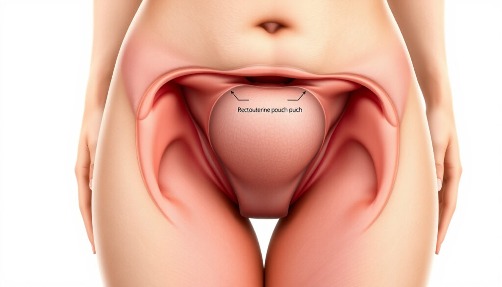
Pouch of Dougla : Anatomy, Symptom , and Condition
The pouch of Douglas is a small peritoneal recess between the uterus and rectum, but its obliteration can signal severe pelvic disease. Endometriosis often targets this space, and ultrasound techniques like the sliding sign help detect it.
Named after Scottish anatomist James Douglas (1675–1742)Location Between the posterior wall of the uterus and the anterior wall of the rectumSignificance Most dependent part of the female peritoneal cavityCommon pathology Endometriosis implants (up to 40% of cases)
Anatomy & Location
- Space between uterus and rectum
- Also called rectouterine pouch
- Deepest point of female pelvis
Common Conditions
- Endometriosis (most common)
- Obliteration (adhesions)
- Fluid collections (abscess, cyst rupture)
Diagnostic Methods
- Transvaginal ultrasound
- MRI
- Laparoscopy (gold standard)
Impact on Health
- Chronic pelvic pain
- Dyspareunia
- Infertility
| Named after | James Douglas, Scottish anatomist (1675–1742) |
| Alternate names | Rectouterine pouch, cul‑de‑sac of Douglas, POD |
| Location | Between uterus (anterior) and rectum (posterior), bounded by uterosacral ligaments |
| Primary function | Allows free movement of organs during bowel and bladder filling; potential space for fluid accumulation |
| Predominant pathology | Endometriosis (up to 40% of affected patients have deposits in the POD) |
| Key diagnostic sign | “Sliding sign” on ultrasound; loss indicates obliteration |
What is the pouch of Douglas?
The pouch of Douglas is a peritoneal recess between the uterus and rectum, also called the rectouterine pouch or cul‑de‑sac Endometriosis Australia. It is named after Scottish anatomist James Douglas (1675–1742) and represents the most dependent part of the female peritoneal cavity.
Anatomical boundaries
Anteriorly lies the posterior wall of the uterus and the supravaginal cervix; posteriorly sits the anterior wall of the rectum. Laterally, the pouch is bounded by the uterosacral ligaments. The deepest point is the Douglas pouch itself, which collects fluid or blood in conditions like ruptured ovarian cysts or ectopic pregnancy.
Clinical significance
Because of its dependent position, the pouch of Douglas is a common site for endometriosis implants — found in up to 40% of affected patients Endometriosis Australia. Obliteration of the pouch can indicate posterior compartment disease PMC pictorial guide. It also serves as a potential space for fluid, making it a target for ultrasound evaluation.
Related medical terms
The pouch is also referred to as the rectouterine pouch, cul‑de‑sac of Douglas, or simply POD. In males, the analogue is the rectovesical pouch between the rectum and bladder.
“The pouch of Douglas is a small area… that has a name and a shape, but the essence of its function remains elusive.” – Dr. T. Sorensen, MJA 2015
What causes pouch of Douglas in females?
The pouch itself is a normal anatomical structure in females. However, pathological conditions can cause it to become abnormal, leading to fluid accumulation, adhesions, or obliteration.
Normal anatomical variation
The depth and shape of the pouch can vary among individuals. A deep pouch may predispose to fluid retention, but this is usually asymptomatic.
Pathological conditions
Endometriosis is by far the most common cause of pouch of Douglas pathology. Inflammation and scarring can lead to adhesions that partially or completely obliterate the pouch PMC pictorial guide. Accumulation of fluid or blood may result from a ruptured ovarian cyst, pelvic inflammatory disease, or ectopic pregnancy.
Risk factors
Factors that increase the likelihood of pouch of Douglas involvement include previous pelvic surgery, history of pelvic infections, and advanced endometriosis. The sliding sign on transvaginal ultrasound can help identify obliteration Step‑by‑step guide to sonographic evaluation of DIE.
Warning: A negative sliding sign suggests adhesions caused by endometriosis AIUM Blog. This finding should prompt further imaging or specialist referral.
What are the symptoms of pouch of Douglas issues?
Symptoms arise when the pouch becomes affected by endometriosis, adhesions, or fluid. The most common presentation is chronic pelvic pain, often with specific triggers.
Pain symptoms
Deep dyspareunia (pain during intercourse) is a hallmark sign. Chronic pelvic pain and dysmenorrhea are also frequent. Site‑specific tenderness at the uterosacral ligaments is commonly encountered and should be documented during ultrasound assessment PMC pictorial guide.
Bowel symptoms
Painful defecation (dyschezia) and altered bowel habits may indicate posterior compartment involvement. These symptoms often worsen during menstruation.
Fertility implications
Adhesions in the pouch of Douglas can impair tubal function and ovulation, contributing to infertility Endometriosis Australia. Obliteration of the pouch is strongly associated with posterior deep infiltrating endometriosis.
“The rectouterine pouch is the most dependent part of the peritoneal cavity in females, making it a common site for fluid collection and seed implantation.” – Radiopaedia editorial
How is the pouch of Douglas seen on ultrasound?
Transvaginal ultrasound is the first‑line imaging tool. The normal pouch appears as an anechoic space posterior to the cervix.
Transvaginal ultrasound appearance
Begin by identifying the hyperechoic posterior pouch of Douglas peritoneum and follow that line carefully PMC pictorial guide. Fluid in the pouch can improve visualization of the uterosacral ligaments and superficial endometriosis.
Free fluid assessment
Free fluid in the pouch may suggest a ruptured ovarian cyst, pelvic infection, or ectopic pregnancy. The amount and character (anechoic vs. complex) provide diagnostic clues.
Signs of obliteration
The sliding sign is used during dynamic ultrasound to assess whether the pouch of Douglas is obliterated Step‑by‑step guide to sonographic evaluation of DIE. Loss of sliding indicates adhesions, often from endometriosis. Obliteration can be described as partial or complete.
The implication: a positive sliding sign effectively rules out pouch obliteration, while a negative sign demands further investigation.
Is there a pouch of Douglas in males?
No — the pouch of Douglas is a female‑specific structure. Males have a different peritoneal recess called the rectovesical pouch.
Male analogue: rectovesical pouch
In males, the peritoneum reflects from the rectum to the posterior wall of the bladder, forming the rectovesical pouch. It is the most dependent part of the male peritoneal cavity and can accumulate fluid or pus.
Embryological differences
The presence of the uterus and cervix in females creates the rectouterine pouch. Without these structures, the male peritoneum takes a different route.
Clinical relevance
Although a true pouch of Douglas is absent in males, disorders of sexual development may produce remnant structures. In practice, the term “pouch of Douglas” is used only for females.
Can the pouch of Douglas affect pregnancy?
Yes — the pouch of Douglas can influence fertility, pregnancy, and delivery, particularly when affected by endometriosis.
Ectopic pregnancy in the pouch
An ectopic pregnancy can rarely implant in the pouch of Douglas, leading to acute abdomen and hemorrhage. This is a surgical emergency.
Adhesions and fertility
Endometriosis‑related adhesions can impair tubal function or trap the ovary, reducing fertility. Even if conception occurs, adhesions may cause pain or complications.
Delivery implications
Severe obliteration of the pouch may complicate cesarean delivery by making the lower uterine segment less mobile. Surgeons must be aware of potential adhesive disease.
Tip: In women with known pouch of Douglas endometriosis, early referral to a maternal‑fetal medicine specialist can help plan delivery.
volusonclub.net, obgyn.onlinelibrary.wiley.com, endometriosisaustralia.org
For a deeper look at how fluid in the pouch of Douglas can affect fertility, explore this detailed guide on pelvic health and anatomy.
Frequently Asked Questions About the Pouch of Douglas
What is pouch of Douglas obliteration?
Obliteration means the normal space is replaced by adhesions or fibrous tissue, often due to endometriosis. It can be partial or complete and is diagnosed by loss of the sliding sign on ultrasound.
Can pouch of Douglas be seen on MRI?
Yes, MRI can visualize the pouch and identify endometriotic nodules or adhesions with high soft‑tissue contrast. It is often used as a complementary tool to ultrasound.
What is the difference between pouch of Douglas and cul‑de‑sac?
They are the same structure. “Cul‑de‑sac” is a French term meaning “bottom of the bag” and is used interchangeably with the rectouterine pouch.
How is endometriosis of the pouch of Douglas treated?
Treatment includes hormonal therapy (contraceptives, GnRH agonists), pain management, and surgical excision via laparoscopy. Complete excision of endometriotic deposits can restore anatomy.
Is pouch of Douglas always present in females?
Yes, it is a normal anatomical feature in all females. However, its depth and patency vary among individuals.
Does pouch of Douglas pain vary with menstrual cycle?
Yes, pain often intensifies during menstruation because endometrial implants in the pouch become inflamed and shed cyclical blood.
The pattern: the pouch of Douglas is a small but clinically vital space; its health directly affects pelvic pain, fertility, and surgical outcomes.
This article also relates to Lower Back Pain Causes and Olympic Park Sports Medicine.
More related posts
 Warwick Meats Product Recall: Mince Beef Plastic Alert
 Kevin Federline Today: Career, Net Worth, Memoir & More
 Palmer Luckey: Net Worth, Biography, Anduril & Controversies
 Village Cinemas Crown – Luxury Recliners and Blockbusters
 Justin Dior Combs: Age, Parents, Career, and Family
 Donald Duck: Real Name, Temper, and Mickey Mouse Comparison
 Lee Jong-suk and IU: Relationship Timeline and Biography
 French Montana: Ethnicity, Net Worth, Sobriety, and More
